News
Discovery of additional neuroimaging evidence for human conscious perception
2022.12.27 Views 1373
Discovery of additional neuroimaging evidence for human conscious perception
Neuroimaging thesis revealing thalamocortical signal looping
The research team led by Prof. Min Byoung-Kyong published its findings in NeuroImage.

Neuroimaging thesis revealing thalamocortical signal looping
The research team led by Prof. Min Byoung-Kyong published its findings in NeuroImage.

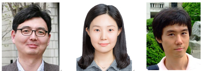
▲ (From left) Prof. Min Byoung-Kyong Min at the Department of Brain Engineering of Korea University, corresponding author; Seo Ji-Hye, research professor at the Institute for Brain and Cognitive Engineering of Korea University, co-first author; and Kim Dae-Jin, a researcher at the Department of Psychological and Brain Sciences of Indiana University, USA (co-first author)
The research team led by Professor Min Byoung-Kyong at the Department of Brain Engineering released its findings that both thalamic inhibitory regulation and thalamocortical integrative iterations are essential for conscious perception. The research team used functional magnetic resonance imaging (fMRI: an imaging method capturing changes in activated brain regions) for this research, and the resulting imaging data corroborated the team’s magnetoencephalography (MEG: measurement of magnetic fields) study in 2020. Both studies were published in NeuroImage, an academic journal (citation index: 7.4) that ranks first in terms of citation index in the field of neuroimaging, and this fMRI neuroimaging study was released online on November 10.
In order to study human conscious perception, the research team designed an experiment to analyze the brain images acquired when participants perceived a fused color of orange after seeing red and green LEDs alternately flickering. During the experiment, only red and green LEDs were presented physically to the subjects and the fused orange color was just cognitively perceived in the mind of participants.
This study, in which Professor Min participated as the corresponding author, was co-authored by Seo Ji-Hye, Research Professor at the Korea University Institute of Brain Engineering and Dr. Kim Dae-Jin at the Department of Psychological and Brain Sciences of Indiana University. It is highly significant that the fMRI neuroimaging of this study and the team’s MEG study of 2020 — representing the two axes of human brain signal study — corroborate the theoretical model hypothesis of thalamus-centered human consciousness announced by Professor Min 10 years ago.
The temporal neurodynamics analysis employed in this study revealed that the difference between the perception of physically lit red and green colors and the perception of the fused orange color only existed in thalamocortical regions related to higher, not lower, internal information processing. This suggests that human conscious perception occurs across higher-order neural networks in the brain, and, furthermore, that signal looping dynamics are necessary for human conscious perception.
Although conscious perception is arguably the highest level of human cognitive function, its neural correlates still remain unclear and thus studying the relationship between the brain and mind has been a long-standing challenge. Professor Min said, “Since the thalamic reticular nucleus (TRN), which plays a key inhibitory role in the human thalamus, is as thin as the skin of an apple, it is not easy to prove its inhibitory function with modern brain imaging technology. However, the two consecutive experiments from the MEG and fMRI studies are significant in that they show that the TRN plays an important role in human conscious perception and that they are the first to demonstrate that human conscious perception is related to the inhibitory action of the thalamus.” This work was supported by the Convergent Technology R&D Program for Human Augmentation, which is funded by the Korean government through the National Research Foundation of Korea.
[ Figure Description ]

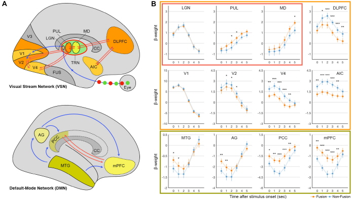
(Figure 1) A. The visual stream network (VSN) and default-mode network (DMN) of the non-fused colors (red/green) and fused color (orange) in conscious perception. Red arrows indicate the thalamocortical area yielding significant differences in brain activity between fusion and non-fusion perception; B. Temporal dynamics of brain activity between the fusion and non-fusion conditions (blue curves: non-fusion; orange curves: fusion; red box: thalamic subdivisions; orange box: VSN; and green box: DMN)

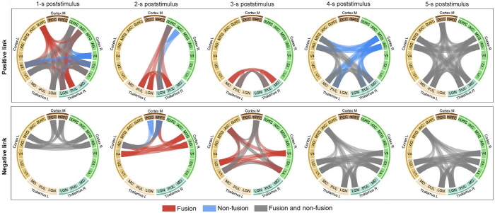
(Figure 2) Correlative graph of the thalamocortical area according to the time of feeling the flickering stimulus (blue: non-fusion perception, red: fusion perception, gray: common perception). At 1-3 s post-stimulus time, there is a difference in the perception of fused and non-fused colors, but this difference disappears at 4-5 s post-stimulus time. At 2 s post-stimulus time, when the most optimal conscious perception occurred, the fusion color showed a positive link with the thalamocortical area.
file

